Understanding Gel Electrophoresis…
What is Gel Electrophoresis?
Gel electrophoresis is a laboratory technique used to separate and analyse molecules, such as large proteins and DNA fragments. This technique uses an electric current to move charged molecules through a gel matrix, allowing separation based on size and charge. It is often used for protein expression, nucleic acid analysis and other molecular biology applications.
DNA and the part it plays in Gel Electrophoresis
DNA (Deoxyribonucleic Acid) is a molecule that contains genetic instructions used in the development and functioning of all living organisms. It is composed of four types of nucleotides, which are arranged in a specific sequence of DNA providing the genetic information for the organism.
During gel electrophoresis, DNA samples are first mixed with a loading buffer that contains a tracking dye, which allows researchers to monitor the progress of the electrophoresis. The DNA sample is then loaded into the wells of the gel, which is usually made of agarose, a material derived from seaweed. Once the gel is loaded with DNA samples, an electrical current is applied. DNA fragments are negatively charged due to the presence of phosphate groups, and they will be attracted towards the positively charged electrode at the other end of the gel. Smaller fragments will travel/migrate through the gel quicker, while larger fragments move more slowly. This separation allows researchers to analyse the size and quantity of DNA fragments in the sample.

Choosing the right Gel Tank
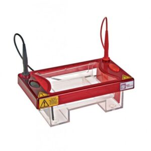
A gel tank is a piece of laboratory equipment used for gel electrophoresis. It consists of chamber that holds the gel and a buffer solution. The chamber has two electrodes, one at each end, which are connected to a power source that generates an electrical field across the gel. The gel is poured into the chamber in a liquid state. Once it solidifies, it forms a matrix through which the DNA/RNA sample can be run. The comb can then be inserted into the gel to create wells, or small pockets, into which the DNA is loaded. The wells ensure that the samples are loaded in a precise and organized manner, allowing for accurate analysis of the separated fragments.
Once the gel is loaded with the samples, the lid of the gel tank is closed, and an electrical current is applied to the electrodes. This causes the charged DNA to move through the gel matrix, with smaller fragments moving more quickly than larger ones. The movement of the fragments is affected by the electrical field and the properties of the gel matrix, which allows for the separation of the samples based on their size.
After the electrophoresis is complete, the gel is removed from the tank and the separated fragments can be visualized using a staining agent, such as ethidium bromide, which intercalates to the DNA and fluoresces under ultraviolet light. The appGLOW Safe Stain from Appleton is ideal to use as it will emit green fluorescence when bound to dsDNA or ssDNA and red fluorescence when bound to RNA. The resulting band pattern can be used to analyse the size and quantity of DNA in the sample, providing valuable information for a variety of research applications.

The ideal product to use to obtain perfect electrophoresis results every time is the multiSUB horizontal gel electrophoresis units by Cleaver Scientific. These gel tanks provide an easy-to-use and flexible platform for all your horizontal electrophoresis requirements. With a wide range of tanks and tray sizes as well as many comb options, the Horizontal Gel Units offers the most versatile solution for DNA and RNA agarose gel electrophoresis currently on the market. The MSMINI, MSMIDI, MSCHOICE, MSMAXI, all offer an unsurpassed combination of gel and buffer volume, with gel size and sample number versatility.

Common issues that can occur during Gel Electrophoresis and how to troubleshoot
There are several common issues that can occur during gel electrophoresis that can affect the accuracy and quality of the results. Here are some examples and potential solutions:
- Smiling or “comet” shape of bands: This can occur when there is too much DNA in the wells or if the voltage is too high. To prevent this, reduce the amount of DNA loaded in the wells or decrease the voltage.
- Poor resolution: This can occur because the wrong buffer was used to make the gel. TAE is used for larger fragments and TBE for smaller fragments OR the concentration of agarose is incorrect. Very small nucleic acids like microRNA need polyacrylamide gels.
- Faint or no bands: This can occur if the DNA sample is degraded or if the buffer solution is not at the appropriate pH. To address this issue, ensure that the DNA sample is not degraded and check the pH of the buffer solution.
Overall, to prevent common issues during gel electrophoresis, it is important to carefully prepare the gel, accurately load the samples, and maintain consistent conditions throughout the electrophoresis process. Troubleshooting problems as they arise can help ensure accurate and reliable results.
Enhance your research results through Innovative Applications of Gel Electrophoresis
There are several innovative applications of gel electrophoresis that can enhance your research results. Here are a few examples:
- Multiplex PCR and gel electrophoresis: Multiplex PCR allows for the amplification of multiple DNA targets simultaneously, which can save time and resources. Gel electrophoresis can be used to separate and analyze the amplified DNA fragments. By combining these techniques, researchers can analyze multiple DNA targets in a single experiment, which can improve efficiency and accuracy
- Plasmids: Gel electrophoresis can be used to excise separated regions of plasmids, both the plasmid backbone and inserts fragment can be covered from the gel for use. By running plasmid DNA samples through gel electrophoresis, it is possible to check the size of the DNA to ensure that the plasmid has been digested correctly. This information can be used to verify plasmid extractions, if DNA has been successfully ligated into plasmids and confirm the presence of specific DNA regions
- RNA electrophoresis: Although gel electrophoresis is most commonly used to separate DNA fragments, it can also be used to separate RNA fragments. RNA electrophoresis can be used to investigate RNA integrity during extraction process and identify if there is any contaminating DNA
Overall, by applying innovative techniques to gel electrophoresis, researchers can enhance their research results by improving efficiency, accuracy, and the depth of information obtained.

Leave a Reply
You must be logged in to post a comment.