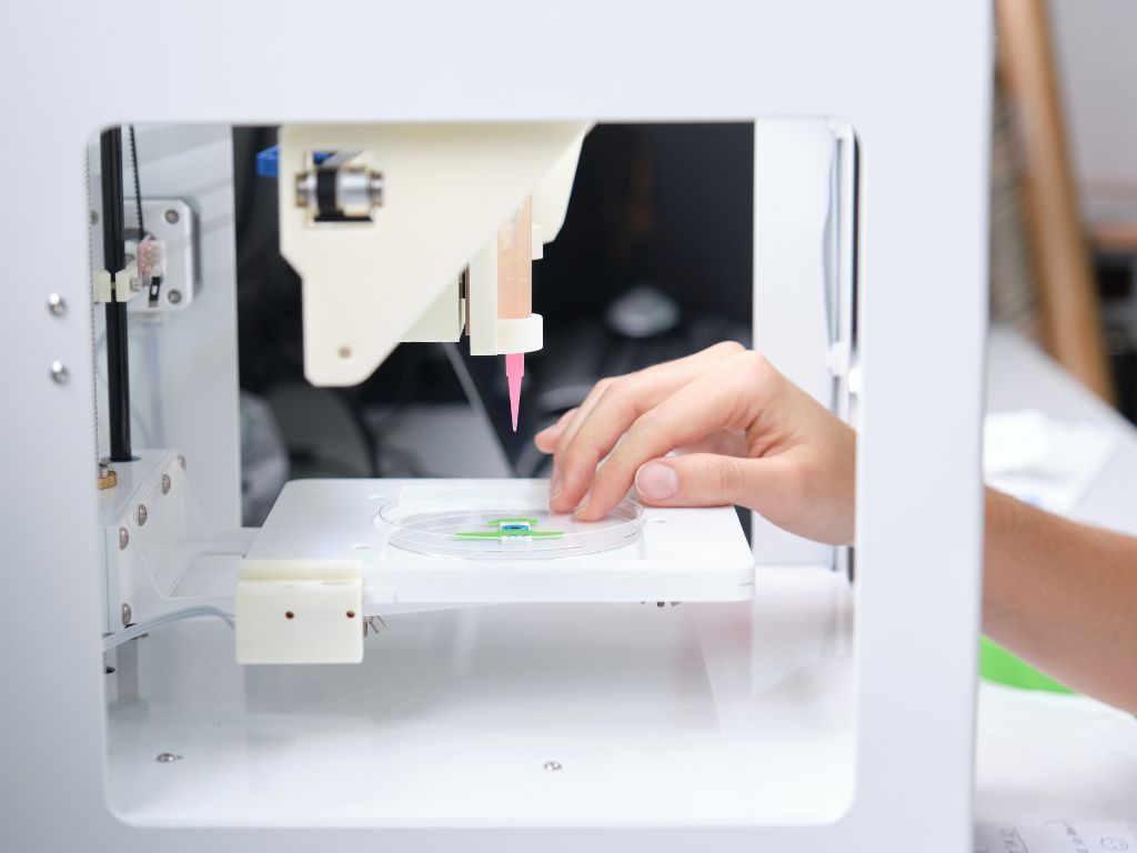
Matribot® Bioprinter- An Easy-to-Use Tool for 3D Cell Culture
Application Notes
By Zhang Linyu and Chen Rui*
Introduction
The Corning Matrigel® matrix was pre-thawed and aliquoted according to the manual. All the tips and syringe used for handling Bioprinting technology has grown along with stem cell research and has become a vital tool with diverse applications in the biological and medical fields.1,2
In vitro 3D cell culture techniques create an accurate in vitro environment and provide an alternative to in vivo models used for fundamental cell biology and physiology research.3
However, these applications are currently hampered by organoid variability, low throughput, and limited scale. Although bioprinting technology is expected to be applied in regenerative medicine and drug discovery, there are enduring concerns regarding the impact of cell dispensers on cell integrity.
The Corning Matribot bioprinter exhibited cell-friendly dispensing performance similar to that of the manual dispensing operation. The culture dispensed by the Corning Matribot bioprinter were suitable for further molecular and protein analysis as well as downstream applications including high-throughput drug screening.
Table 1. Printing parameters for dispensing cell suspensions and mouse intestinal organoid fragments
Materials and Methods
Bioprinting with cell suspensions and intestinal organoid fragments
The Corning Matrigel® matrix was pre-thawed and aliquoted according to the manual. All the tips and syringe used for handling spheroids or organoids were pre-chilled. The mediums were equilibrated to room temperature (15°C to 25°C) before use.
A549 and MDCK cells were cultured in Dulbecco’s Modified Eagle’s Medium (DMEM; Corning 10-013-CV) supplemented with 10% Fetal Bovine Serum (FBS; Corning 35-081-CV) until they reached 90% confluence.
The medium was removed by aspiration, and the cells were washed with Phosphate-Buffered Saline (PBS; Corning21-040-CV) and dissociated into single cells using 0.25% TrypsinEDTA (Corning 25-053-CI). An equal volume of complete medium (DMEM + 10% FBS) was then added to the cells and mixed.
The suspension was centrifuged at 300 x g for 3 min., the supernatant was discarded, and the pellet was resuspended in undiluted Corning Matrigel® matrix (Corning 356231). The final cell density was adjusted to 50 cells/µL. The mixture of the Matrigel matrix and cells were transferred to a syringe (BD 309657), inserted into the printhead of the Corning Matribot bioprinter (Corning 6150), and dispensed using the parameters listed in Table 1.
Manual dispensing was performed using a single channel pipettor. The plates were incubated at 37°C for 15 min., followed by the addition of complete medium (1 mL) to each well. The plates were incubated at 37°C and 5% CO2, and the medium was changed every 2 to 3 days. Mouse intestinal organoids (MIOs; STEMCELL Technologies 70931) were thawed, cultured, and passaged in complete IntestiCult™ organoid growth medium (STEMCELL Technologies 06005), according to the manufacturer’s instructions, until a density of approximately 150 organoids/well was achieved.
A pre-wetted 1,000 µL pipet was used to break up the organoids by pipetting up and down 20 times, followed by centrifugation at 290 x g for 5 min. at 4°C. The supernatant was carefully discarded, and the pellet was resuspended in undiluted Matrigel matrix by pipetting up and down 10 times.
The mixture of Matrigel matrix and MIO suspension was transferred to a syringe, inserted into the printhead of the Corning Matribot bioprinter, and dispensed using the parameters listed in Table 1.
Manual dispensing was performed using a single-channel pipettor. The plates were incubated at 37°C for 15 min., followed by the addition of complete IntestiCult organoid growth medium (1 mL) to each well. The medium was changed three times per week.
Click here to view product details
Immunohistochemical analysis of 3D cell cultures
As described in the Guidelines for Use (CLS-AN-528)4, A549 and MDCK spheroids or MIOs were collected from Matrigel matrix using Cell Recovery solution (Corning 354253), washed several times with cold PBS, and fixed with 4% paraformaldehyde at 4°C overnight. The 3D cultures were washed with PBST (PBS + 0.05% Tween® 20) and permeabilized with 0.2% Triton™ X-100 for 30 min. The 3D cultures were washed with PBST several times prior to staining.
For immunostaining, the 3D cultures were incubated overnight at 4°C with primary antibodies (Table 2) at 1:100 dilution. The next day, the 3D cultures were washed with PBST, incubated with fluorescent secondary antibodies, and the nuclei were stained with 2 µg/mL 4′,6 diamidino-2-phenylindole (DAPI). Images were captured using a CQ1 Image cytometer.
Table 2. Antibodies for immunofluorescence
Table 3. Primers for gene expression analysis
Gene expression analysis
After dissociation from the Matrigel matrix, the 3D cultures were washed several times with cold PBS (Corning 21-040-CV). RNA was extracted using a Magnetic Tissue/Cell/Blood Total RNA kit(TIANGEN DP761). One-Step TB Green® PrimeScript™ RT-PCR kit (Takara Bio RR066A) with primers synthesized by GENEWIZ (Table 3) was used with a LightCycler® system (Roche) for real-time quantitative polymerase chain reaction (qPCR) analysis.
Results and Discussion
We demonstrated that 3D cultures dispensed by using the Corning® Matribot® bioprinter were comparable with those obtained via manual operation. Both methods had brought similar morphology, component cell types, and gene expression profile.
As illustrated in Figure 1A, the morphology of A549 spheroids generated from bioprinter-dispensed and manually dispensed samples seemed to have normal shapes.
After culturing for 7 days, the bioprinter-dispensed and manually dispensed MDCK spheroids showed obvious and typical cystic structures (Figure 1B). Complex, multilobed MIO structures were also observed after 5 days of culture (Figure 1C). Moreover, the densities of the formed 3D cultures were comparable between the bioprinter-dispensed and manually dispensed samples (Figures 1A-C).
Immunofluorescence staining was performed to investigate the presence of specific cell type markers in the 3D cultures. Significant junctional F-actin was observed at the cell-cell contact points within the A549 spheroids, indicating that stable and strong intercellular adhesive interactions were formed.5
Moreover, 3D cultures of A549 cells showed bright and continuous E-cadherin labelling on the cell surface and at cell-cell contact sites. Hence, immunofluorescence staining confirmed that F-actin and E-cadherin were highly expressed in A549 spheroids generated by both bioprinting and manual dispensing (Figure 2A). Töyli M, et al.6 reported that when MDCK cells are cultured in a 3D environment, they form cell cysts with the apical domain facing a lumen, and with E-cadherin delineating the lateral membranes and ZO-1 at the tight junctions within the lumen. In agreement, the present experiment showed that MDCK spheroids generated by both methods expressed E-cadherin and ZO-1 (Figure 2B).
Moreover, immunohistochemical analysis of the MIOs cultured using bioprinted and manual methods showed that the 3D cultures contained differentiated intestinal cells, including enterocytes, goblet cells, and Paneth cells, as demonstrated by the expression of Villin, Mucin-2, and Lysozyme, respectively (Figure 2C), which was consistent with a previous report.7
In accordance with Liu J, et al.,5 the lipopolysaccharide receptors CD14, MD2, and TLR4, were upregulated in 3D cultures as compared with 2D cultures. Herein, the levels of these receptors in both bioprinted and manually generated spheroids were comparable and higher than those in monolayer cell cultures (Figures 3A-C).
The formation of MDCK spheroids was previously reported to be accompanied by a reduced expression of the apoptosis inhibitor Survivin and of the vesicle transport effector Rab7, but with increased Rab5 expression.6
Consistent with these findings, herein Rab5 was also found to be upregulated, whereas Rab7 and Survivin were downregulated in MDCK cultures compared with 2D cultures (Figure 3D-F). In addition to remarkable proliferative activity, the cellular composition of the intestinal epithelium is extremely diverse, reflecting a high degree of heterogeneity within its major lineages.8 In line with the intestinal epithelium cellular nature, qPCR analysis confirmed that the experimental MIOs expressed the coding genes of Lgr5, Villin, Vimentin, TFF3, Lysozyme, and chromogranin-A and -B, which are specific to intestinal stem cells, enterocytes, mesenchymal cells, goblet cells, Paneth cells, and enteroendocrine cells, respectively (Figure 3G).
Noteworthy, a high level of consistency of gene expression profiles was observed between 3D cultures generated by the Corning Matribot bioprinter and manual dispensing were comparable between the bioprinter-dispensed and manually dispensed samples (Figures 1A-C).
Immunofluorescence staining was performed to investigate the presence of specific cell type markers in the 3D cultures. Significant junctional F-actin was observed at the cell-cell contact points within the A549 spheroids, indicating that stable and strong intercellular adhesive interactions were formed.5 Moreover, 3D cultures of A549 cells showed bright and continuous E-cadherin labelling on the cell surface and at cell-cell contact sites.
Figure 1. Representative photomicrographs of A549 and MDCK spheroids, and mouse intestinal organoids (MIOs). Brightfield of manual generated and bioprinted 3D cultures. (A) A549 spheroids after 10 days in culture. (B) MDCK spheroids after 7 days in culture. (C) MIOs after 5 days in culture. Images were captured at 40X and 100X magnifications with an Olympus IX53 microscope.
Figure 2. Immunohistochemical staining of specific cell types in the 3D cell cultures. Representative photomicrographs of fluorescently stained (A) A549 spheroids, (B) MDCK spheroids, and (C) mouse intestinal organoids. Images were captured at 200X magnification with an YOCOGAWA CQ1 Image Cytometer.
Figure 3. Gene expression profile in A549 and MDCK spheroids, and mouse intestinal organoids (MIOs). (A) CD14, (B) MD2, and (C) TLR expression in A549 monolayer and spheroid 3D cultures. (D) Rab5, (E) Rab7, and (F) Survivin relative mRNA expression in MDCK monolayer and spheroid 3D cultures. (G) Cell -specific maker expression in MIOs. Data represent mean ± SD of experimental triplicates.
Conclusion
3D cell cultures generated using the Corning® Matribot® bioprinter were similar to those obtained by manual dispensing in terms of morphology, component cell types, and gene expression profile.
The culture dispensed by the Corning Matribot Bioprinter were suitable to be analysed by various techniques, including observation under a microscope, immunohistochemical analysis, qPCR, etc.
If you have any questions on the Corning® Matribot® Bioprinter email us here.
1. Murphy SV and Atala A. 3D bioprinting of tissues and organs. Nat. Biotechnol. 32, 773–785 (2014).
2. Peng W, Datta P, Ayan B, et al. 3D bioprinting for drug discovery and development in pharmaceutics. Acta Biomater. 57:26-46 (2017).
3. Jiang T, Munguia-Lopez J, Flores-Torres S, et al. Bioprintable alginate/ gelatin hydrogel 3D in vitro model systems induce cell spheroid formation. J. Vis. Exp. 137:57826 (2018).
4. Extraction of three-dimensional structures from Corning Matrigel matrix guidelines for use (Corning, CLS-AN-528).
5. Liu J, Abate W, Xu J, et al. Three-dimensional spheroid cultures of A549 and HepG2 cells exhibit different lipopolysaccharide (LPS) receptor expression and LPS-induced cytokine response compared with monolayer cultures. Innate Immun. 17:245-255 (2011).
6. Töyli M, Rosberg-Kulha L, Capra J, et al. Different responses in transformation of MDCK cells in 2D and 3D culture by v-Src as revealed by microarray techniques, RT-PCR and functional assays. Lab Invest. 90: 915-928 (2010).
7. Sato T, Vries RG, Snippert HJ, et al. Single Lgr5 stem cells build cryptvillus structures in vitro without a mesenchymal niche. Nature. 459:262-265 (2009).
8. Norkin M, Capdevila C, Calderon RI, et al. Single-cell studies of intestinal stem cell heterogeneity during homeostasis and regeneration. Methods Mol. Biol. 2171:155-167 (2020).


